White pines are one of the most numerous native trees in this area.
Yet, it's sometimes difficult to determine whether or not they have white canker infections.
Since I've noticed a number of white pines whose needles are turning brown and dropping off,
I decided to check a few out.
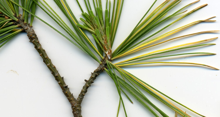
The following white pine was in decline, and probably dying.
This sample branch was taken and analyzed in early June.
1
As you can see here, there are some green needles, and some that have turned yellow and died.
But unlike other tree species, there are very few spores along the branch or at the branch junction.
2
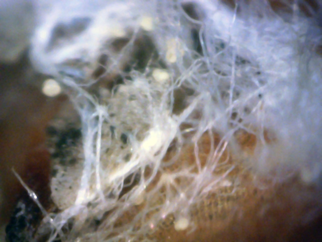
The telltale spores and hyphae are actually there, but difficult to see.
As shown in this 400x microscopic view, the hyphae appear as "cotton-like" tiny areas at the point where the 5-needle packets erupt from the bark.
The spores are scattered throughout the hyphae.
3
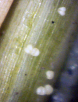
These spores were on the base of a needle. (400x)
I returned to this tree in early November, after the old needles were shed and the new needles
were created. I snipped a branch and put it under the microscope. The needles visually looked fine
and there was no evidence of white canker when looking at their cross-sections. Likewise, very few spores
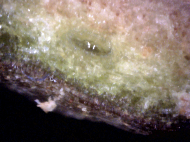
were found on the surface of the twig bark. Finally, I examined the cross-section of a twig.
The results are shown below.
4
↑
↑
↑
↑
↑
Look closely and you can see a diffuse blob of white canker in the upper-left corner.
Also, the lower-right corner shows another more concentrated blob of white canker
that is shooting a runner toward the bark, then under it, then breaking the surface,
then creating a blob above the surface.
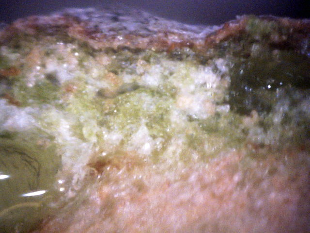
5
A small side branch was pruned off, and the scar on the larger host branch was
examined in this picture. Note the canker's preference for rich green tissue,
and the diffuse nature of the white canker growth.
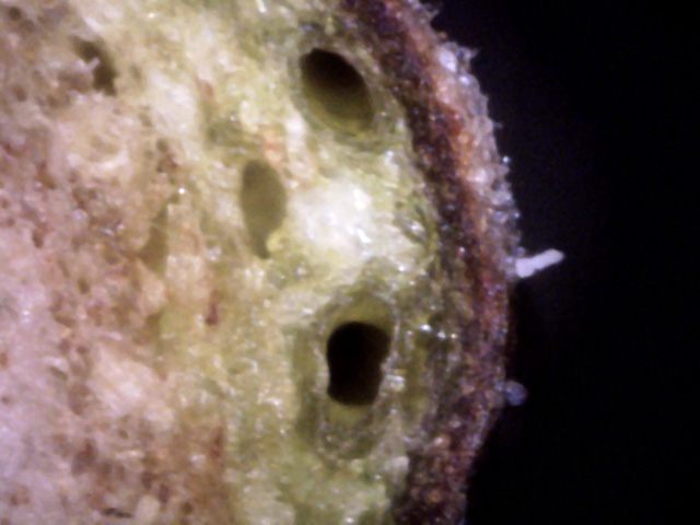
6
Two things of note here:
1) The white canker grows near but avoids the large vessels.
2) The blob of white canker between the vessels is very likely the source of the white canker area on the bark near it.
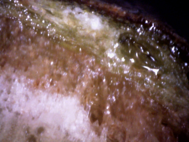
7
White canker in the outer cortex, and at the xylem-phloem junction.
The proximity is probably more than a coincidence.
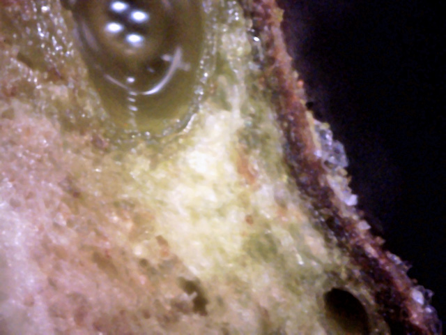
8
Once again, the canker growth is near a conducting vessel, but does not enter it.
And this canker area has spawned some translucent canker on the bark surface.
White canker appears to have a difficult time infecting white pine trees.
The needles and bark show little or no evidence of an infected state.
While microscope views at 400x of twig cross-sections will reveal the white canker, its appearance
is often diffuse, and it clusters near the sap transporting vessels. But something
in these vessels or sap keeps the white canker from attacking them.
This is very different from deciduous trees, where white canker seems to know no bounds.
Hence, it appears that white pines will stand up to a white canker infection longer than
deciduous trees
I located another nearby large white pine whose needles were also half gone due
to disease, and obtained a sample branch from it. These pictures were taken in
early June, just about the time of a major spore release. The analysis results are shown
below.
 ↓
↓
1
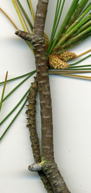
This is a branch from that diseased tree. It fits the
typical profile of white canker: moderate spore density scattered on the branch
bark with a high concentration at branch and leaf junctions that are on the
bottom of the branch (red arrow).
2
3
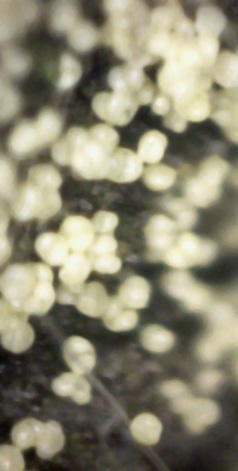
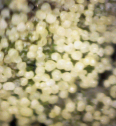
Here are two pictures of the yellow spore area pointed at by the red arrow in
picture 1. They were made using a 400x digital microscope.
4
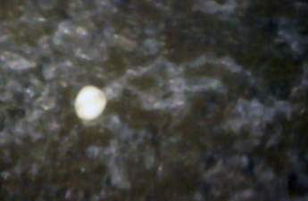
This is a single spore and its likely associated hypha just under the Bark. This
picture was also taken at 400x.
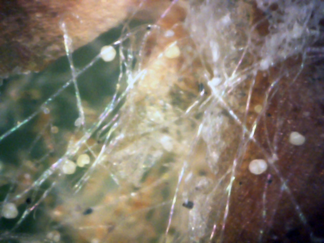
5
Here is another 400x view of an area of the bark that contains both spores and hyphae.
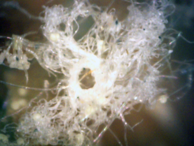
6
This appears to be a massive hyphae tangle along with a number of spores
in the background. This is a 400x view.
I went back to this same tree in early October to get another branch for testing, with the
intent of doing a more in-depth analysis using needle and twig cross-sections using
a digital microscope. We begin by looking at the surface of a needle from a 1/8 inch twig.
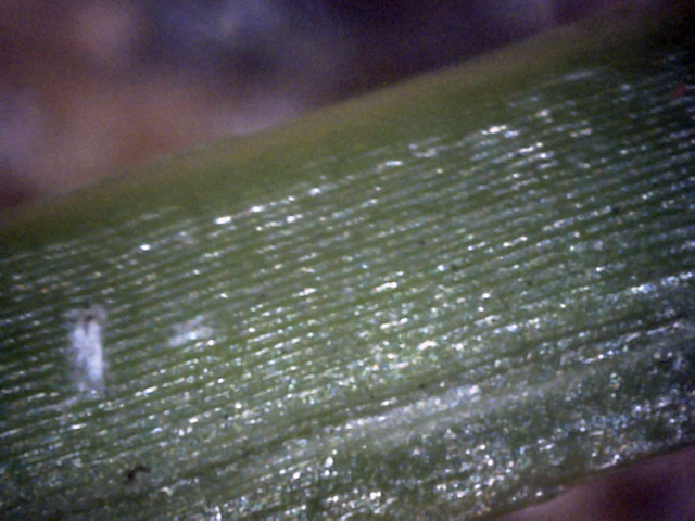
7
Here is a plain, generic needle surface.
Only tiny pieces of white canker evidence are present, like at the center-left,
and they would probably be easy to overlook.
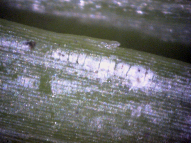
8
This needle surface shows more white streaks of material that is probably white canker.
Interestingly, near the top it almost looks like overexposed words!
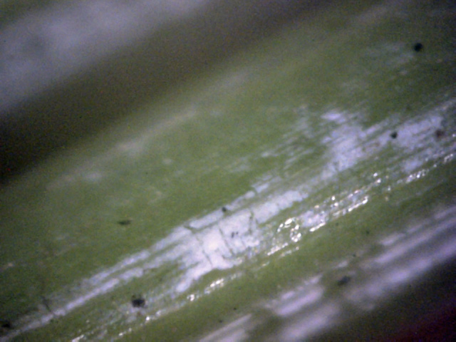
9
This is probably a white canker infection on the needle surface, but it almost
resembles snow because of its diffuse pattern.
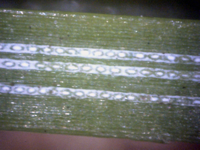
10
The stomata on the surface of a needle.
It's unclear if the white stomata and the white surround are actually due a white canker
infection, since I haven't found any closeup views of healthy white pine needles
to compare against.
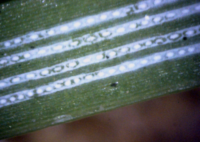
11
These stripes seem to be common on many needles.
But they aren't always as uniformly spaced as shown here.
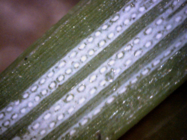
12
Prior pictures have shown these stomata in separate rows.
Here two rows are grouped together. That may be just a chance occurrance.
But I'm guessing that the white stomata and the white surround are due to
white canker, although the evidence isn't as strong as I haven't seen
this infection pattern on other needles or leaves.
The following set of 400x microscopic pictures shows cross-section views of several needles.
Like the prior surface pictures, there is some evidence for white canker, but it isn't strong.
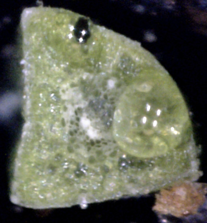
13
Not much canker infection shown here.
What little there is seems to be on the outside of the needle and within the vacuoles
at the needle center.
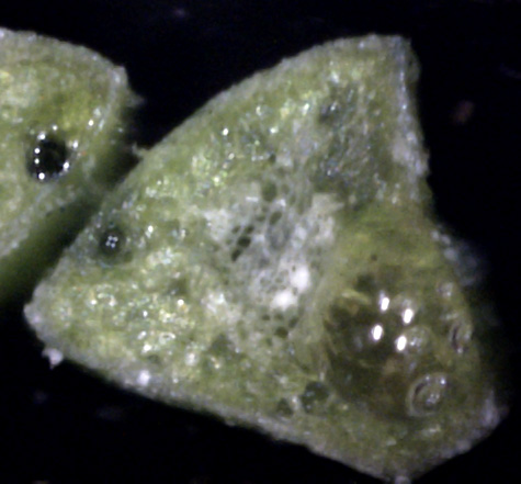
14
This picture shows a bit more canker infection at the needle's surface and a bit
more within the center of the needle.
But the needle doesn't otherwise appear badly infected.
This agrees with the outward appearance of the needle, which was normal in appearance.
Part of another needle is shown in the upper left.
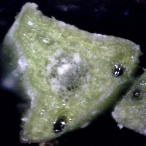
15
Of the needle cross-sections shown, this may be the one showing the worst infection.
But once again, the infection sites are the same - the needle surface and the pith
at the center of the needle. And once again, big blobs of white canker seem to be absent
- all the infection seems to be diffuse.
When needle and leaf surface views and cross-sections aren't very helpful for providing
good evidence for white canker, twig cross-sections can be very informative, as shown in
the following microscopic views at 400x.
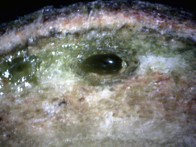
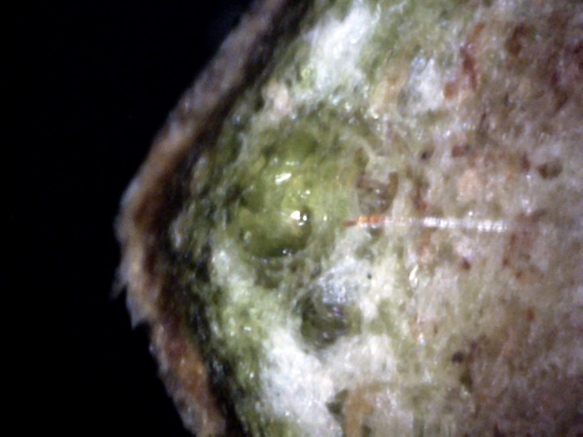
16
Lots of dispursed white canker growth in the phloem, and a big bundle around the
conducting vessel. The bark seems to be riddled with diffuse canker.
Interestingly, the sap in the vessel seems to ward off the white canker.
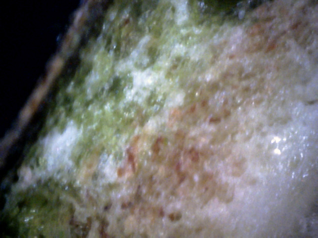
17
More evidence of white canker.
Also, again note the tendency of the canker to congregate at the outer phloem and near
the conducting vessel, where the energy supply is richest, i.e., food is plentyful.
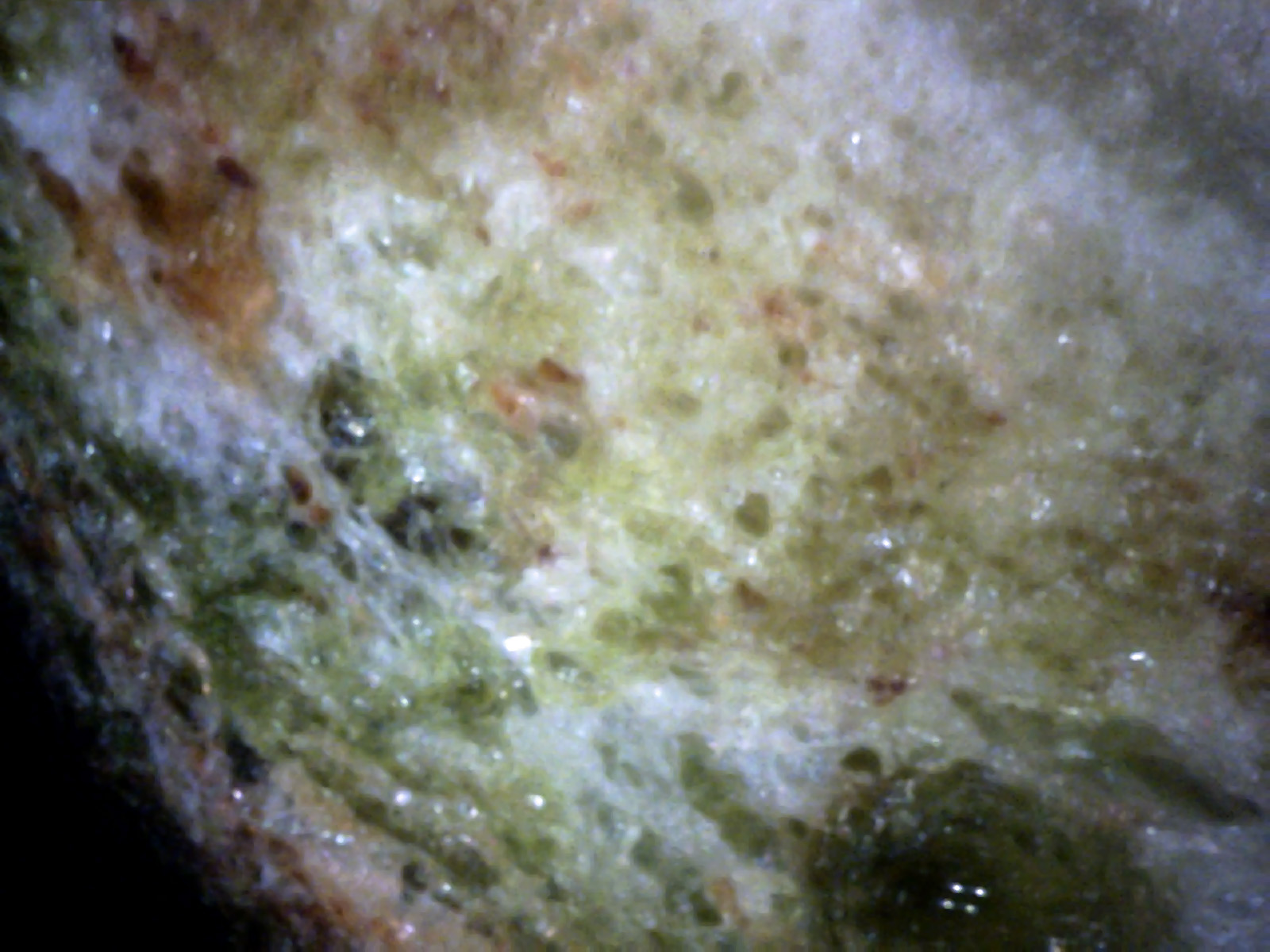
18
Not all white canker is located near the vessels, as this picture shows.
Also evident is that the dark green cortex is hard to find - there is so much white canker
mixed in with it.
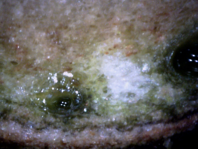
19
There is so much diffuse cloud-like white canker in this area that it almost looks
like a work of art.
In fact, this may be the prettiest white canker picture of all the trees and shrubs
I've examined!
20
This is a great example illustrating that white canker tends to seek out areas under
the bark where the food supply is at its maximum - within the phloem
and near the vessels (but not too close to the sap, which it seems to avoid).
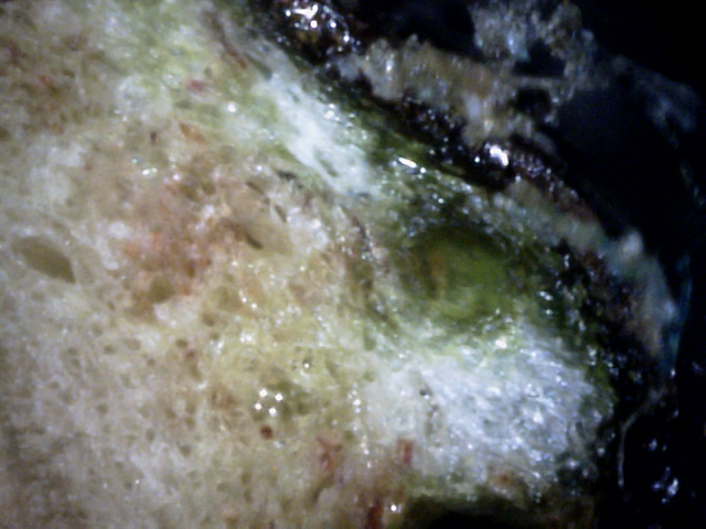
21
Another area of intense white canker growth, again next to a vessel.
Missing is the appearance of more distinct blobs of white canker.
The closest we come to blobs seems to be shown here, in or on the bark surface,
where grayish blobs are seen.
In summary, it appears as if white pines definitely can be infected with
white canker. However, evidence for this infection is difficult to see when
examining either the surface or cross-section of a needle. (White highlighting of needle
stomata lines may also be an indicator.)
The best evidence for white canker is available in May and early June, when
white canker spores are present at certain locations on the branches, and can be
examined microscopically. The most reliable year-around
evidence is obtained by examining the cross-section of a twig microscopically
using a 400 power microscope, where the white canker shows itself as diffuse
white patches in the outer phloem, adjacent to the conducting vessels.
While on a trip to the south shore of Lake Superior in Northern Michigan,
I saw quite a few trees in decline.
I clipped off a branch of a white pine that was half-dead and brought it home for analysis.
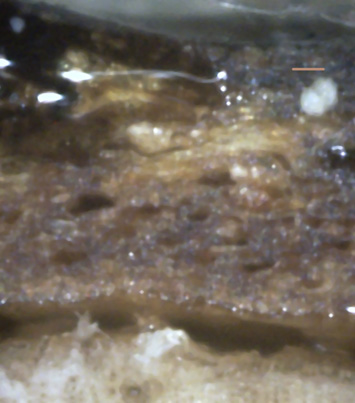
I chose to examine a cross-section of a branch that was 3/16 inch thick.
 ↑
↑
↑
↑
↑
↑
1
I found little evidence of infection in the inner bark.
But the outer bark was a different story.
The yellow arrow in Picture 1 shows a mature spore buried within the bark.
The beige line above it is a 50μm scale bar.
Note that there is also some white waxy substance growing between the inner
and outer bark (green arrows).
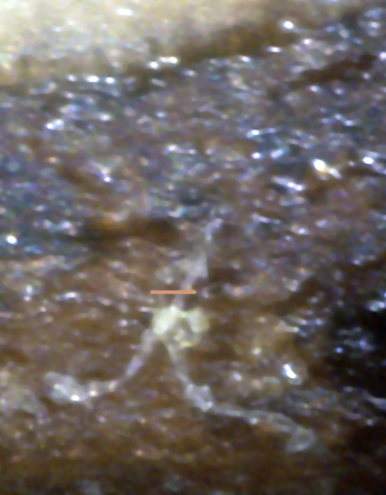
2
Picture 2 shows another mature spore and hyphae within the bark.
A 50μm scale bar is also shown alongside it.
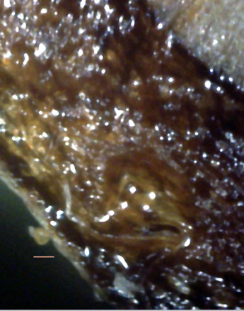
3
→
←
←
↓
Finally, here we see a young spore growing on the outside of the bark (yellow arrow).
A hyphae tangle lies underneath it (green arrows), and a single hypha appears to be growing
from the young spore.
In summary, this small but important tree analysis shows that even as far away as
Wisconsin, white canker is attacking trees there the same as it is here in New England!


